Date: December 12, 2020
Best,
Brendan Stack, MD Vice Chair AHNS Endocrine Section
Greg Randolph, MD Chair AHNS Endocrine Section
Advancing Education, Research, and Quality of Care for the Head and Neck oncology patient.
Published on by AHNS Webmaster
Best,
Brendan Stack, MD Vice Chair AHNS Endocrine Section
Greg Randolph, MD Chair AHNS Endocrine Section
Published on by AHNS Webmaster
Instructional Course: Management of Salivary Malignancy Case Based Panel Discussion
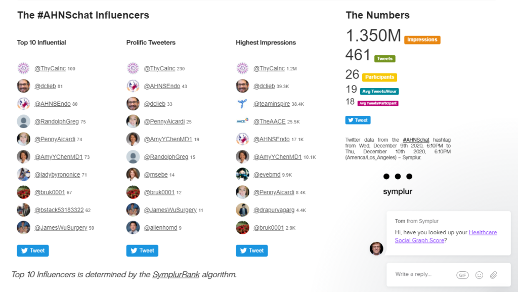
Date: Wednesday, December 9, 2020
Time: 4:00 PM Pacific /6:00 PM Central / 7:00 PM Eastern
Session is an hour long
Complimentary to all attendees
At the conclusion of this webinar, participants will be able to:
Faculty:
Moderator: David Cognetti, MD, Thomas Jefferson University
Panelists:
Cecelia Schmalbach, MD MSc FACS, Temple University
Kiran Kakarala, MD, University of Kansas
William Ryan, MD, University of California, San Francisco
Published on by AHNS Webmaster
The American Head and Neck Society will sponsor nine research grants for 2021 which are available through American Academy of Otolaryngology CORE. The Letters of Intent (LOI) for the Grants are due by 11:59 EST on Tuesday, December 15, 2020.
You may find general information about the AHNS grants by visiting the website: https://www.ahns.info/research/grants/
Applicants interested in applying for the AHNS grants must submit their applications through the Academy CORE (combined otolaryngologic research evaluation) process. Letters of intent and applications are paperless and submitted through proposal Central.
Reminder: You must complete the Letter of Intent before submitting the full grant. You will not be penalized if you submit an LOI and decide not to complete a full grant.
Published on by AHNS Webmaster
Moran Amit, Hideaki Takahashi, Mihnea Paul Dragomir, Antje Lindemann, Frederico O Gleber-Netto, Curtis R Pickering, Simone Anfossi, Abdullah A Osman, Yu Cai, Rong Wang, Erik Knutsen, Masayoshi Shimizu, Cristina Ivan, Xiayu Rao, Jing Wang, Deborah A Silverman, Samantha Tam, Mei Zhao, Carlos Caulin, Assaf Zinger, Ennio Tasciotti, Patrick M Dougherty, Adel El-Naggar, George A Calin, Jeffrey N Myers.
From Nature, February 2020; 578(7795):449-454.
Article Review by Moran Amit, MD, PhD and Patrick J. Hunt, MD, PhD
Background / Hypothesis
Solid tumors are complex collections of cells that are encircled by benign cells that both influence and are influenced by the tumor. These surrounding cells, known as the tumor microenvironment, include vasculature, immune cells, neurons, and other cell types. Tumors manipulate these nearby cells in ways that benefit the tumor. Autonomic neurons have been shown to innervate and drive malignant growth in a variety of solid tumors, including head and neck, prostate, breast, and gastric cancers. However, the mechanisms by which these neuron-tumor relationships are formed are not well understood. In this study, we hypothesized that the trophic relationships between oral cavity squamous cell carcinomas (OCSCCs) and nearby autonomic neurons arise through direct signaling between the tumors and local neurons.
Design
This study was conducted using several laboratory models. OCSCC progression was modeled in mice by genetic knockout of the Trp53 gene (encoding the p53 protein) combined with administration of the carcinogen 4-nitroquinoloine 1-oxide (4NQO), which mimics the genetic alterations associated with tobacco use. Additional studies were conducted in cell culture models in which p53-deficient cells were co-cultured with mouse trigeminal ganglia neurons. Xenograft models were also used, in which OCSCC cells were injected directly into the oral cavities of mice. Using these models, we examined changes in the cellular composition of the tumor microenvironment and in changes in microRNA expression in cancer cells, and the effects that these changes had on the tumors and associated neurons.
Summary of Results
Loss of p53 expression in OCSCC tumors resulted in increased nerve density within the tumors. This neuritogenesis was controlled by shuttling of tumor-derived microRNAs via extracellular vesicles. The microRNAs within these extracellular vesicles were primarily responsible for regulating the observed axonogenesis. Of these vesicular microRNAs, miR-34a acted s a major restrictive factor for neuritogenesis, whereas miR-21 and miR-324 activity promoted neural reprogramming and neuritogenesis. Moreover, the neurons innervating p53-deficient OCSCC tumors acquired adrenergic features through transdifferentiation of trigeminal sensory nerve fibers. This transdifferentiation process corresponded with increased expression of neuron-reprogramming transcription factors, including POU5F1, KLF4, and ASCL1, which are overexpressed in p53-deficient samples and are candidate targets of miR-34a regulation. In OCSCC samples from human patients, increased expression of tyrosine hydroxylase, a marker of adrenergic neurons, was strongly associated with poor outcomes, thereby demonstrating the relevance of these findings in human patients with cancer.
Strengths
Weaknesses
Key Points
From the Basic Science/Translational Service
Jeffrey C. Liu MD Vice Chair
Richard Wong MD Chair